
Model : BTH-300S Probes : 2 Unit Price (USD) : 32,500 Certificate : CFDA Remark : Full digital wave-beam imaging system Multi-beam parallel processing technology THI Imaging Pulse inverse harmonic imaging Spectrum noise rejection Dynamic focusing technology Color flow peak velocity capture Color suppression imaging DirPower(Directional Power) Doppler flow imaging Continuous wave Doppler imaging Tissue Doppler imaging Wideband, multi-frequency probe imaging The following message should be added to each productâ??s description: Minimum Order Size :1 Product origin : China Self-manufacture
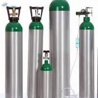
This recertified Medical MD Oxygen Cylinder is perfect for professional or personal home oxygen use. With a 14.6 cu ft capacity,this medical oxygen cylinder is perfect for portability and smaller spaces. This lightweight, easy to transport aluminum requalified cylinder is DOT approved, and ready for medical or industrial use. It comes fitted with a CGA870 post valve installed and a freshly painted dome in a Medical Green color for easy identification. PREMIUM CONSTRUCTION Reconditioned High-quality aluminum body Clearcoat protected Medical green rust-resistant painted dome CGA870 medical post valve installed Approximately 4.4â?³ in diameter x 16.3â?³ in height* (with valve) Weighs approximately 6 pounds Capacity 14.6 cubic foot 2015 PSI service / 3360 PSI test pressure Stamped DOT-3AL2015 5-year retest interval Recertified test within 180 days prior to shipment
Material: BAMBOO. Product Type: FAN. Use: Holiday Decoration & Gift. Regional Feature: Viet Nam. Place of Origin: Vietnam. Brand Name: 99 GOLD DATA. Model Number: Selena +84 587 176 063. Logo: Fan Cover. Product name: Bamboo Fan Bamboo Hand Fan. Color: Natural Bamboo Color. Usage: Promotional Gift. Design: Classic. Feature: Natural Bamboo Handmade.
Product Details: Model Number 6200 Brand BBS Connectivity Type Wired Type of operation Handheld Color Black Scan Speed 55 Scan Lines Per Second Backed by in-depth industry experience, we are involved in manufacturing and supplying an extensive range of high quality Scanners.
Summary of main specifications and system of laptop 4D color Doppler color Doppler ultrasound Laptop type all digital color Doppler ultrasound host Ultrasonic host operating system: Windows7 operating system Spectrum pulse Doppler Direction energy Doppler Real time three synchronization Space composite imaging: the requirement is 3 level, visual adjustable. Organized harmonic imaging technology 4B imaging mode One key intelligent optimization Support multilingual user interface Monitor: 15 inches, high definition LED Physical clipboard: save the image on the left side of the screen, which can be deleted directly. The system has the function of on-the-spot upgrade Presupposition: for different inspection of the viscera, preset the inspection conditions for the best image, reduce the adjustment of the operation, and the commonly used external adjustment and combination regulation. Support real-time 3D imaging function Language: Chinese/English/Russian/Spanish/French/Arabic/Vietnamese/Portuguese/Indonesian The probe interface is 1
APPLICATION: hospitals, clinics, health care centers, universities and etc . Examination range: 3D, abdominal, obstetrics, gynecology, regional nerve block, uterus / adnexa, vascular and etc .
Features for Ultrasound Scanner: DBF: Digital Beam Forming RDA: Real-time dynamic aperture imaging DRA: Dynamic real-time acoustic apodizer DRF: Dynamic receiving focus DFS: Dynamic frequency scanning Ensure that the image is not distorted, edge clearer, level richer Humanized operating design 8 sections TGC gain adjustment With abundant built-in software packages Comprehensive software measurement capabilities, to allow more extensive clinical application Two probe connectors Probe connected with a variety of choice to achieve maximum functionality. Excellent value for money VLSI, advanced technology, stable performance Reasonable cost control, to achieve the perfect embodiment of price and performance Specification for Ultrasound Scanner: Image magnification: *0.8, *1.0, *1.2, *1.5, *1.8, *2.0 Local zoom: 2 times local zoom in real time Dynamic range: 64dB â?? 96dB adjustable Focus: 4-segments dynamic electronic focuses selected Gray scale: 256 Pre-processing: variable aperture, image direction, dynamic filter, edge enhancement, etc. Post-processing: digital space time filter, 8 y corrections, 16 Pseudo Colors, column correlation, frame correlation, spot correlation, linear interpolation, etc. Multi-frequency: 2.5MHz/3.0MHz/3.5MHz/4.0MHz etc. Multi-frequency selected Measurement: distance, circumference/acreage, HR, pregnancy week (BPD, GS, CRL, FL, AC) and calculating fetus weight, etc. Annotation: Chinese/English interface transition; hospital name, doctors/patients name, case number, gender, age; 16 body marks with probe location, full screen character annotation, real time clock display Puncture lead: Image B: Puncture guide line under B mode Gain adjustment: 8-segments TGC adjustment, GAIN adjustment or near field, far field, overall gain adjustable Image reverse: left/right, black/white, up/down Storage: 128 images permanent storage Cine loop: 256 images real time display cycling/one-by-one checked Output interface: 2 SVGA video outputs, SVGA color monitor circumscribed; 2 PAL video outputs, which can be connected with PAL standard monitor, Video thermal recorder, ultrasound image workstation, etc. Standard configuration: 12.1 inch SVGA high resolution monitor 2 probe connector 1 USB Optional: 80 elements R60, 3.5MHz multi-frequency convex probe High frequency linear probe Electronic trans-vaginal probe Rectal probe Video thermal recorder 3D Ultrasound image working station, etc. Package Content: - Main Unit: 1 each - Power cable: 1 each - Ground Line: 1 each - Fuse (3A): 2 each - User Manual in English: 1 each - Packing list: 1 each - Optional part: 1 set
Applications: Cows, horses, pigs, sheep, alpacas, rabbits, dogs, cats, foxes, snakes, Fish and other animals. Description: This equipment is convex linear ultrasound scanning diagnostic system with high resolution. It applies micro-computer control and digital scan converter (DSC), digital beam-forming(DBF), real time dynamic aperture (RDA), real time dynamic receiving apodization, real time dynamic receiving focusing(DRF), digital frequency scan (DFS), 8 segments digital TGC, frame correlation technologies to endue its image with clarity, stability and high resolution.
Boxianglai Bxl-V10 Equine Ultrasound Scanners Farm Vet Ultrasound Machine for Sale Description: The BXL-V10 Veterinary Ultrasound Scanners are mainly used to test the pregnancy of animals, estimate the gestational age, the number of births, and uterine diseases, and can also detect symptoms such as empty pregnancy and stillbirth. Commonly used in major breeding farms, etc. Scope of application Suitable for pregnancy diagnosis of animals such as swine, horse, bovine, sheep, etc. Specifications: 1. Model Name: BXL-V10 4.Display 5.6 HD LCD Screen (640 480 Pixel) 5.Imaging Modes: B / 2B / 4B / BM / M 6. Image Gray Scale: 256 7. Host Storage: 64 frames 8. Cine Loop: 400 frames 9.Scan Depth Adjustment: 70mm-240mm 10.Image Flip Function: up/down left/right 12. Image Processing: Pseudo-color 15.Measurement: Distance Circumference Area Volume 16.Gestational: Age Expected birth date 17.Characters and comments: Date Time Full-screen character editing 19. Continuous Working Time: 3.0 Hours 20. Battery Capacity: 3000mAh 21. The structure of the device Standard Kit (1)Host device (Include build-in Li-On battery) (2)3.5MHz Convex Probe (CXA50R) (3)Power adapter (Including power cable) (4)User manual Optional Accessories (1)6.5MHz Rectal Linear Probe (LNA64) (2)5.0MHz Micro-convex Probe (CXA20R) (3)3.5MHz Convex Probe (CXA50R) (4)7.5MHz Linear Probe (LNA40)
Application OB/GYN Abdomen scan Superficial parts Urology Ophthalmic scan MSK Nerve block Feature Integrated clipboard, display the saved image at the bottom of the screen, which can be directly called or deleted Preset optimized image inspection conditions for different inspection organs, reducing adjustment procedures during operation Abdomen, uterine attachment, heart, fetus, urinary system, prostate, blood vessels, small organs, newborn, musculoskeletal, intraoperative scan Complete Probe Family,� probe box silicone lining, effective protection of probe Subarray Technology Harmonic Imaging Technology (THI) CE certified
Basic Info. Model NO. BW-5 Gain up to 120dB Pseudo Color 16 Colors Selectable N.W, 6kg Power Supply AC 110-220V 50Hz Connectors Two Transducer Connector Cine Loop 256 Frams Scanning Depth up to 250mm Image Memory up to 128 Frames Trademark BENEMED Transport Package Carton with Foam Inside Specification 48*38*41cm Origin China HS Code 9018121000 Product Description Customer Question & Answer BENEMED BW-5 Portable Ultrasound Diagnostic Scanner Portable B/W Ultrasound Scanner with Clear Image Quality Feature: Digital Beam Forming Superior image quality Dynamic Receiving Apodization Dynamic Frequency Scan Advanced imaging processing USB port for convenient image transfer Portable B/W Ultrasound Scanner with Clear Image QualityPortable B/W Ultrasound Scanner with Clear Image Quality Specification General Description: Imaging Modes: B, B/B, B/M, 4B, M Scanning Modes: Convex, Linear Gray Scale: 256 Display: 12'' LED monitor Gain: up to 120dB Scanning dept: up to 250mm Imaging technology: Digital Beam-forming Dynamic Frequency Scan Dynamic Receiving Focusing Dynamic Receiving Apodization Real-time Dynamic Aperture Imaging Processing Pre-processing: 8-segment TGC adjustment Dynamic range Frame correlation Edge enhance Post-processing: Gray scale Polarity reverse Left/Right reverse Up/down reverse Functions: ZOOM: Local zoom Panoramic zoom Image memory: 128 frames Cine Loop: 256 frames View: Patient info, patient image, Patient report Pseudo color: 16 colors selectable Software Functions General measurement: distance, circumference, area, volume, angle, ration, etc Calculation package: abdomen, cardiac, gynecology, obstetrica, urology, orthopedics, small parts, etc Obstetric measurement: BPD, CRL, GS, FL, HC, AC, EDD, AFI, FW Comments: Week, day, time, doctor time, patient ID, age, sex, transducer type, transducer frequency, gray scale Peripheral ports: USB SVGA PAL-D Video output Others: Net weight: 6.0KG Power supply: AC 220V±22V 50Hz Dimensions: 480*380*410 Standard Configurations: BW-5 main unit 12'' LED monitor Two transducer connectors 256 frames cine loop Electronic convex transducer Options: Electronic linear transducer Electronic endocavity transducer High frquency rectal linear transducer Mobile trolly Thermal printer
Dolphi C full digital color Doppler System ultrasound meditech Free hand 3D imaging - Multi-imaging modes: B/C/M/PW/PDI/Triplex - High-precise digital continuous beam formation - Dynamic Frequency Fusion Imaging Technology - High-precise delay point-by-point dynamic receiver focus Ultra-wide band imaging technology - Adaptive image optimization processing technology - THI tissue harmonic imaging technology - Precise SRI adaptive speckle noise suppression #Colordoppler #Doppler #ultrasound_Scanner #Ultrasound #Meditech #china
8 Inch Veterinary Ultrasound Scanner Bxl-V50 for Sheep/Pig/Cow/Bovine/Equine 1. Durable Veterinary Ultrasound with IP67 dustproof and waterproof; 2. Large capacity rechargeable Li-On battery for extended working time (6-7 hours) 3. Multi kinds of probes could be chosen, for Bovine / Sheep / Goat / Swine / Camel..... 4. Perfect imaging quality with large storage memory Features 1. 8.0inch HD screen with 800*600 pixel 2. Perfect imaging quality without vague 3. More than 40 kinds of animal body remarks support 4. Scan Frame Rate for 60 frames/s with Cine loop function support 5. More than 8 kinds of grid settings and quick measurement support Warranty for 1 year
General Specification 1. Main Features � Operationg SyetemWindow 7 Monitor 12.1 inch LED high-resolution 90� rotatable monitor Scanning ModeB, B/B, 4B, B/M, M, 3D Image Reconstruction KeyboardBack-lit Silicone Keyboard (option) Cine LoopUp to 1024 frames Gain0-255dB TGC8 segment Scanning DepthUp to 310 mm Dynamic Range20-150dB Pseudo-color40, or customized Body Marking140 Focus4 Magnification0.8-6.0 Probe Connector2 LanguageChinese/English/Spanish Matching PrinterLaser/Vedio/Inkjet/Thermal printer Peripheral PortUSB, DICOM3.0, RS232, VGA, VCR, PAL-D Measurement & CalculationB-mode: Distance, Perimeter, Area, Volume, Angle, Histogram, Cross Section, Hip Joint Angle M-mode: Heart Rate, Time, Gradient Image Processing TechnologiesTHI, Pre-process, Post-process, Dynamic Range, Contrast, Frame Correlation, Average Line, Edge Enhancement, Black-white Change over, Gray-scale Transformation&Adjustment, Brightness, Gamma Correction � 2. Applications Abdominal(Liver, Gallbladder, Spleen, Kidney, Pancreas) Gynecology(Uterus, Cervix Uterus, Endometrium, Ovary, Ovarian Follicle, Hydatoncus, Enclosed Mass) Obstetric(AC, BPD, CRL, FL, THD, GS, OFD, HUMERUS, TIDIA, ULNA, HC, TAD, APTD, FTA, AFI, Analytic report of fetus and the growth curve) Cardiology(LV, LV Function, Aorta, Mitral, Valve) Urology(Left Renal Volume, Right Renal Volume, RUV, Prostate kidney) Small Parts(Ophthalmology, Thyroid, Jaw and Face) 3. Standard Configuration Main system unit, 3.5MHz Convex probe, Power adapter, User manual and disc 4. Optional Items 4 hours backup lithium battery, Biopsy Bracket 5. Probes Selection 3.5 MHz Convex Probe (3.5-14MHz variable frequency ) 7.5 MHz Linear Probe (3.5-14MHz variable frequency ) 7.5 MHz Transvaginal Probe (3-10MHz variable frequency) 5.0 MHz Micro-Convex Probe (2-6MHz variable frequency) 7.5 MHz Rectal Probe(80 elements) 6. Advanced Technologies PC platform Full Digital Ultrasonic Kernel Engine� One Touch Optimizer Tissue Harmonic Imaging 3D Imaging Reconstruction DICOM 3.0 Packaging & Delivery Packaging Details: Double Box Package� Net Weight: 4.5kg� Gross Weight: 8kg Package Size: 460*400*310mm Delivery Time: 5-10 working days Warranty Two years for main unit (One year warranty for probes) Please let us know your specific request. We would like to recommend you the most suitable model based on your information.
Kaon Series Ubiquitous OCT Scanner Portable optical coherence tomography (OCT) scanner for ophthalmology Product Specifications: - Center wavelength: 840 nm - A-scan rate: 26,000 per second (typical) - Imaging depth: 0.9 mm (in air) - Output power: 750 - Depth resolution: 9 micrometers (in air) - DC adapter: 24 V, 5.0 A output - Lateral resolution: 17 micrometers - Dimension: 240 mm(W), 94 mm(D), 380 mm(H) - Pixel resolution: (volume) 480x120, 200x200, (B-scan) 1024x21, 1024x19, 480x9 circle - Weight: 4.5 kg - Scan range: 6.5mm x 6.5mm - Display Size: 15.6-inch Full HD touch Panel
Canyearn C75 Full Digital Trolley Ultrasonic Diagnostic System Color Doppler Ultrasound Scanner General Specification 1. Main Features � Operationg SyetemPC Windows platform Monitor 15 inch LED high-resolution monitor with rotatable arm Scanning ModeB, 2B, B/M, 3D/4D Dynamic Range0-150dB Cine LoopUp to 1024 frames Gain0-100dB TGC8 segment Scanning DepthUp to 300mm Magnification2.0 - 10 Body Marking63 Probe Connector4 LanguageChinese/English Matching PrinterLaser/Vedio/Inkjet/Thermal printer Peripheral PortUSB, LAN, S-VIDEO, RS232 Blood Flow ImageCW, CFM, PW Normal MeasurementDistance, Area, Angle, Volume, Trace, Diam red(diameter stenosis), Area red(area stenosis), Cross curve, Parallel lines M General: ET, HR, Vel, Dis D General: ET, HR, Vel, ACC, Common, Trace Professional MeasurementAbdominal, Cardiology, Urology, Vascular, Early Obstetric, Later Obstetric, General, MSK, Pelvis Small organs(right/left thyroid) Combination of Digital Ultrasonic TechnologyThe multi-core intel processor The wideband digital beam forming The multi beam parallel processing Trapezoid Imaging Accurate blood flow imaging Intelligence speckle suppression Wide view imaging Intelligence screen capture One touch optimization � 2. Standard Configuration Main system unit, 3.5MHz Convex probe, 7.5 MHz Linear Probe, Image storage unit, Specialist report, Image processing optimization, User manual and disc 3. Optional Items 3D+4D, Biopsy Bracket, USB disk 4. Probes Selection 3.5 MHz Convex Probe (3.5-14MHz variable frequency ) 7.5 MHz Linear Probe (3.5-14MHz variable frequency ) 7.5 MHz Transvaginal Probe (3-10MHz variable frequency) 5.0 MHz Micro-Convex Probe (2-6MHz variable frequency) 7.5 MHz Rectal Probe(80 elements) 3D/4D Probe Cardiac Probe 6. Advanced Technologies PC platform One Touch Optimizer Tissue Harmonic Imaging 3D/4D Imaging Reconstruction DICOM 3.0 Packaging & Delivery Packaging Details: Wooden Box Package� Net Weight: 70kg� Gross Weight: 79kg Package Size: 700*790*930mm Delivery Time: 5-10 working days Warranty Two years for main unit (One year warranty for probes)
Canyearn c95 plus full digital built-in ultrasonic diagnostic system light trolley color doppler ultrasound scanner with touch screen General specification 1. Main features  Operationg syetempc windows platform Monitor 15 inch led high-resolution monitor with a touch screen Scanning modeb, m, c, d Cine loopup to 1024 frames Gain0-100db Tgc8 segment Scanning depthup to 240mm Magnification1.5-5.0 Body marking60 Probe connector4 Languagechinese/english Matching printerlaser/vedio/inkjet/thermal printer Peripheral portusb, vga, dicom, rs232 Blood flow imageb, m, c, d Normal measurementm mode: hr, slope, time, dist b/c mode: vascular D mode: velocity, auto trace, hr, accel, pi, ri, time, ps/ed, m trace Professional measurementabdomen(liver, cbd, gb, portal vd diam, spleen, panc, aorta diam, kidney), gyn, urology Combination of digital ultrasonic technology�?�the multi-core intel processor �?�the wideband digital beam forming �?�the multi beam parallel processing �?�trapezoid imaging �?�accurate blood flow imaging �?�intelligence speckle suppression �?�wide view imaging �?�intelligence screen capture �?�one touch optimization 2. Standard configuration Main system unit, 3.5mhz convex probe, 7.5 mhz linear probe, image storage unit, specialist report, image processing optimization, user manual and disc 3. Optional items 3d+4d, biopsy bracket, usb disk 4. Probes selection 3.5 mhz convex probe (3.5-14mhz variable frequency ) 7.5 mhz linear probe (3.5-14mhz variable frequency ) 7.5 mhz transvaginal probe (3-10mhz variable frequency) 5.0 mhz micro-convex probe (2-6mhz variable frequency) 7.5 mhz rectal probe(80 elements) 3d/4d probe Cardiac probe 6. Advanced technologies Pc platform One touch optimizer Tissue harmonic imaging 3d/4d imaging reconstruction Dicom 3.0 Packaging & delivery Packaging details: wooden box package� Net weight: 60kg Gross weight: 69kg Package size: 700*790*930mm Delivery time: 5-10 working days
Features of pl-2018ci digital portable ultrasound scanner: 1. Microcomputer and digital scanning converter are equipped in this digital portable ultrasound scanner. And its images are very clear, stable and high-resolution. 2. Four display modes have been designed for it: b, b+b, b+m, and m. Its grayscale is 256. 3. It can freeze, and enlarge real-time images, and it can also reverse its images up and down, left and right, and black and white. 4. It has 10 kinds of different body marks, and lithotriptor positioning function is also available. 5. Its display includes fixed characters, time and date. 6. The electronic focusing mode is employed, and its focuses can randomly combine with each other. 7. Built-in pseudo-color processing module, and pal-d and vga video output have been designed. 8. It can work together with large-screen monitor, color displayer, video recorder, video printer, and more. 9. Touch keyboard and mouse operation makes it easy, convenient and flexible to use. 10. It is compact in assembly, small in size, and light in weight. 11. Its standard configuration is 3.5 mhz multi-frequency convex probe, and its optional configurations include 7.5 mhz linear probe, 6.5 mhz transvaginal probe, and mitstubishi p93 thermal video printer.
Features: 1.Sleek and fully backlighted panel 2.2 probe connectors 3.8-segment TGC 4.10inch high-resolution monitor 5.Image-Memory and storages function 6.Complete Calculation package for OBSTETRICS, GYNECOLOGY&CARDIAC.
Technical Specification: Scanning mode: convex / linear / micro-convex Probes: 96 elements convex probe, trans-vaginal probe, trans-rectal probe, high frequency linear probe Monitor: 15inch high resolution LED Operation panel: user-friendly, convenient and flexible with back-lit silica gel keyboard & track ball operation Display mode: B, B+B, B+M, M, 4B Detecting depth: 242mm Resolution: lateral 2mm, axial 1mm Blind Zone: 3mm Geometry precision: lateral 5%, axial 5% Gray scale: 256 Electronical focusing: 2 focuses combination randomly Cine loop: 256 frames, realized in automatic successive playback and manual single step forward, backward playback USB port: storable and readable Image storage: 8 Local zoom can be used at real time. Magnification: 0.8, 0.8, 0.90, 0.95, 1.0, 1.05, 1.10, 1.20, 1.30, 1.40, 1.50, 1.60,1.80, 2.0, 2.2, 2.5 (16 kinds) Body marks: 30 Probe interface: 2 Pseudo color: 7 Image processing: up/ down, left / right, black / white conversion, edge enhancement, gamma correction, frame correlation, pseudo color processor (need external color display). Measurement: distance, perimeter, area, volume, heart rate, gestational week, FW, AFI, EDD, maternity table, heart package etc. Note: date, time, name, sex, age, doctor, hospital, comment, Chinese/ English conversion. Output interface: PAL-D, SVGA (SVGA color monitor), USB port. â?¢ With the puncture guiding function, line position can be adjusted â?¢ With gravel positioning, dynamic target area tracking function Main power supply: AC220V±22V, 50/60Hz Probe convex probe trans-vaginal probe trans-rectal probe high frequency linear probe Flexible and convenient operating system: 15inch LED liquid crystal display, angle range is more flexible to adjust Back-lit keyboard silicone gel, more comfortable and wearable, no trouble in using in the darkroom Intelligent menu, man-machine dialogue is easy and quick Puncture guide line can display under B mode, angle and position are adjustable More magnification display, Disease diagnosis more accurate USB storage is connected outside, more convenient to upload pictures Large capacity of cine loop, image can display circularly and automatically


